Integrity, Compassion, Striving for Excellence
Ultrasonography
Ultrasonography is an essential and highly versatile diagnostic tool in modern equine practice. It allows us to examine soft tissues in real time, providing invaluable information to support accurate diagnosis and effective treatment planning.
While ultrasound is most commonly used for orthopaedic assessment, particularly of tendons, ligaments, and other soft tissue structures, it is also widely used to examine internal organs including the liver, intestines, kidneys, spleen, lungs, and eyes. It also plays a vital role in equine reproductive work.
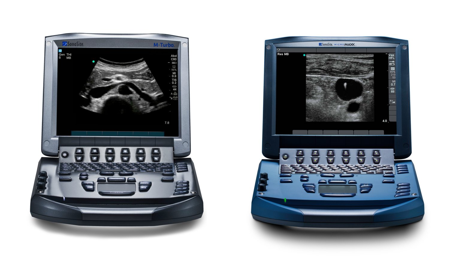
We have invested heavily in advanced ultrasound equipment to ensure we can obtain high-quality diagnostic images without the need for hospital referral in most cases. Our practice is equipped with a range of modern scanners, including high-resolution systems for detailed orthopaedic imaging, alongside versatile portable units for abdominal, thoracic, and ophthalmic examinations.
Advances in ultrasound technology in recent years have significantly improved image quality and diagnostic capability, allowing us to assess structures with greater accuracy than ever before.
Need More Information?
If you would like to discuss whether ultrasonography may be beneficial for your horse, please contact the practice - our team will be happy to advise.
-

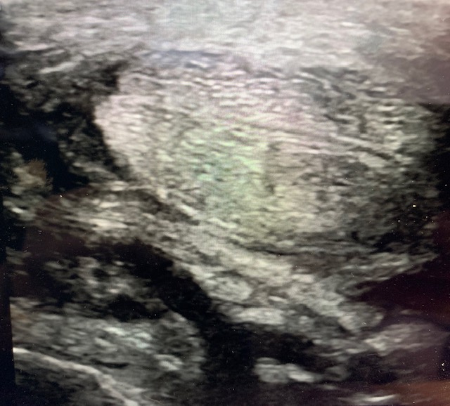
Tendon scan showing flexor tendons and check ligament
-
-
What Can We Assess?
Ultrasonography is an extremely versatile tool and our portable ultrasound machines are highly adaptable and capable of delivering excellent image quality across a wide range of clinical situations.
We have invested in two brand new LogiQ ultrasound scanners that provide the most detailed pictures for orthopaedic imaging. We use them for evaluation of tendon and ligament injuries, including fibre disruption, enlargement, and healing progress; assessment of muscles and some joint structures such as cartilage (e.g. within the stifle)
Our Draminski-Blue scanner is ideal for assessment of abdominal organs such as the liver, spleen, and kidneys, as well as intestinal thickness and motility in cases of colic, weight loss, or diarrhoea. It also allows thorough investigation of respiratory disease, by evaluating the thorax (chest) of both adult horses and foals. We can also visualise the internal eye structures when direct examination is not possible
Detailed trans-rectal imaging of the ovaries and uterus allow us to monitor reproductive cycles, assist with timing of insemination, perform pregnancy diagnosis, and investigate fertility or hormonal issues
We also have two wireless handheld scanners that provide fast and effective decision making in colics.
Most of our ultrasound equipment is fully portable and battery powered, allowing us to perform detailed examinations at your yard. This flexibility enables prompt, accurate diagnosis with minimal stress and disruption for your horse.